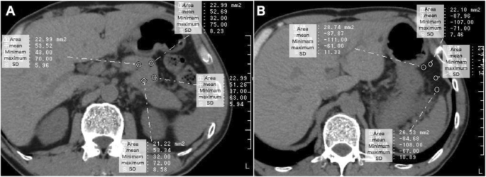
Scan time 116 min. Transabdominal ultrasound us and magnetic resonance imaging mri are commonly used for the examination of the pancreas in clinical routine.

Liver Measurement Ultrasound Pancreas And Its Proportions
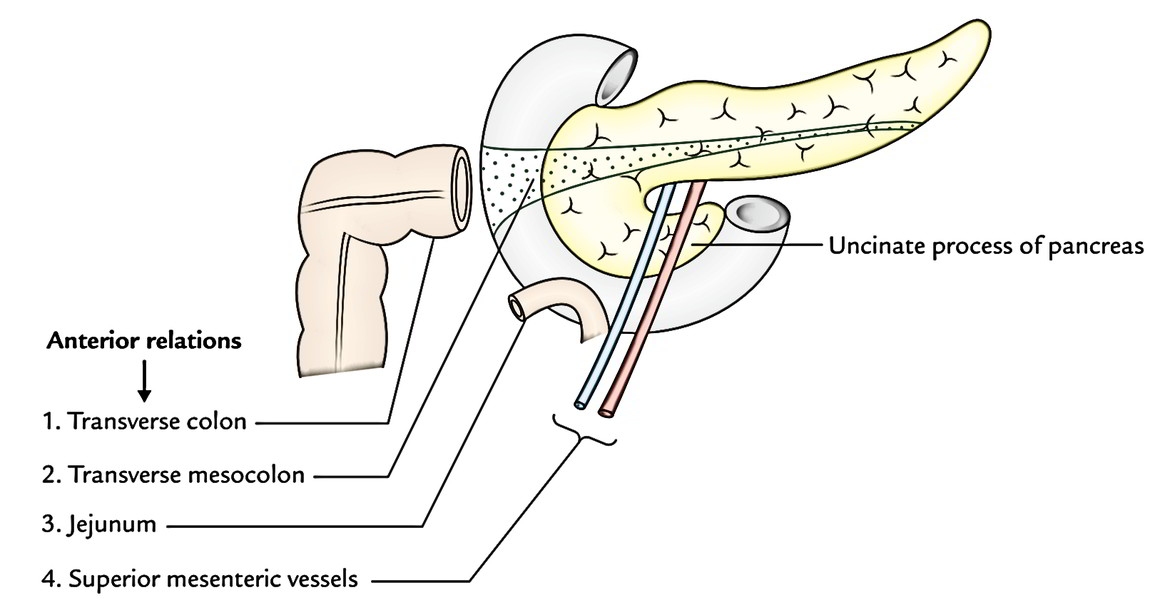
Pancreatic body measurements. And the possible causes of misinterpretation of the pancreatic axial tomography are considered. The middle sections are the neck and body. Pancreatic cysts are diagnosed more often than in the past because improved imaging technology finds them more readily. Approximate normal measurements are. The pancreas is a gland about six inches long located in the abdomen. The wide end of the pancreas on the right side of the body is called the head.
Many pancreatic cysts are found during abdominal scans for other problems. 12 20 cm pancreatic duct. In humans the average pancreas volume was 72745 ml range from 350 to 1055 ml. This result is in strong agreement with results of previous large postmortem and computed tomography ct studies. In assessing these values it is important to be sure that adjacent structures such as the. After taking a medical history and performing a physical exam your doctor may recommend imaging tests to help with diagnosis and treatment planning.
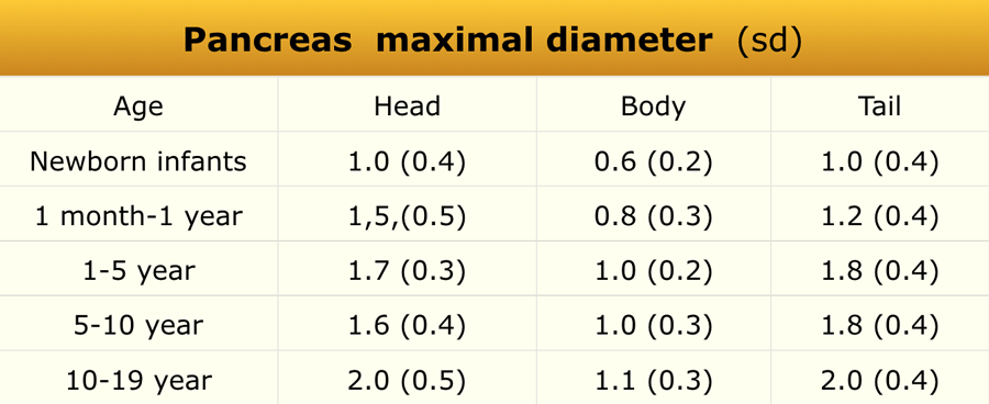
The size of the normal pancreas was found to be up to 30 cm for the head 25 cm for the neck and body and 20 cm for the tail. Downstream tumor or stricture. Pixel size 23 16 mm gap size of 12 mm. Pancreatic size was measured using an axial fat saturated t1weighted flash 2d sequence acquired with 60 mm slice thickness and the following imaging parameters. In mini pigs the measurements of pancreatic volume by mri and by water displacement were almost identical r2 09867. The pancreas is about 6 inches long and sits across the back of the abdomen behind the stomach.
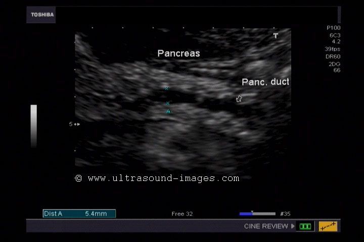
Head 35mm anterior to posterior neck 10 15mm tail 20mm. Abnormal dilatation of the pancreatic duct indicates obstruction of the normal flow of pancreatic secretions due to a distal ie. Its normal reported value ranges between 1 35 mm 58. The diameter of duct can increase with inspiration 3. We therefore were interested in the concordance of these two imaging methods for the size measurement of the pancreas and how age gender and body mass index bmi affect the organ size. The head of the pancreas is on the right side of the abdomen and is connected to the duodenum the.
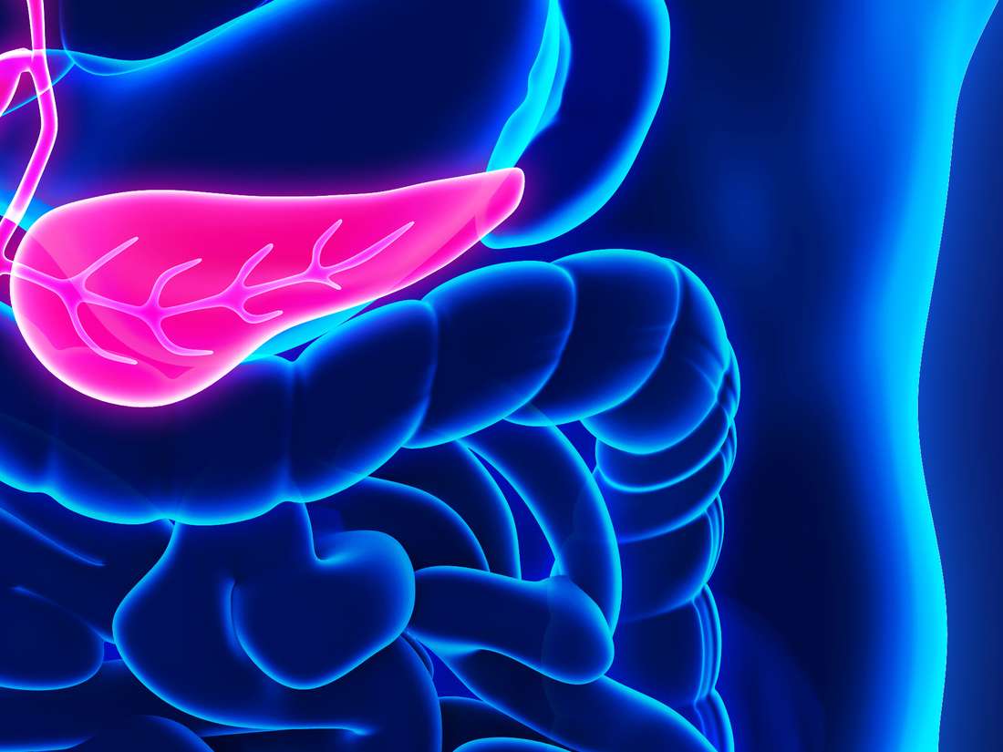
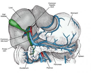
It is shaped like a flat pear and is surrounded by the stomach small intestine liver spleen and gallbladder.
